Today I want to tell you about a basic part of our body.
That part is most important, without that we cannot do any work properly.
That part is our eyes.
The eye isour organ of sight. The eye has several components that include but are not limited to.
Our eyes have many parts.
Part of our eyes:-
i) Cornea
ii) Sclera
iii) lens
iv) Choroid
v) Retina
vi) Iris and Pupil
vii) Ciliary body/Ciliary muscle
viii) Suspensory ligament
ix) Fovea centralis
x) Optical disc (Blindspot)
xi) Blood Vessels and Optic nerves
These are the parts of the eyes.
Functions of the parts:-
i) Cornea:-
The cornea is your eye's clear, protective outer layer. Along with the sclera (the white of your eye), it serves as a barrier against dirt, germs, and other things that can cause damage. If your cornea is damaged by disease, infection, or an injury, the resulting scars can affect your vision.
If the damage to your cornea can't be repaired, doctors can remove the damaged part and replace it with healthy corneal tissue from a donor. Artificial cornea. As an alternative to corneal transplant, doctors can replace a damaged cornea with an artificial cornea, called a keratoprosthesis
ii) Sclera:-
The sclera is the white part of your eye. It's a tough, protective covering and the muscles that control eye movement are connected to it.
The white part of the eye, called the sclera, is a protective layer that covers more than 80% of the eyeball's surface. A healthy sclera is white.
Long-term use of certain medications can give your sclera a blue-gray tint. Certain antibiotics, such as minocycline used to treat rosacea and rheumatoid arthritis can produce this effect. Minocycline can also discolor the skin, ears, teeth, or fingernails, making them appear blue-gray.
iii) lens:-
The lens of the eye is a biconvex, relatively acellular, optically transparent intraocular structure that with the cornea serves to transmit light to the retina with minimal light scattering. Early anatomists viewed the lens as the location of meaningful visualization.
The lens of the eye, also called the crystalline lens, is an important part of the eye's anatomy that allows the eye to focus on objects at varying distances. It is located behind the iris and in front of the vitreous body. It's also flexible, so it can change shape and bend the light to focus properly on the retina.
The crystalline lens of the eye is a natural lens that produces one-third of the eye's total optical power and focuses light into an image on the retina (the light-sensitive tissue at the back of the eye). The crystalline lens is elastic which allows it to flex to change its shape.
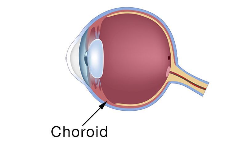
iv) Choroid:-
The choroid is the middle layer of tissue in the wall of the eye. It's found between the sclera (the whites of the eyes) and the retina (the light-sensitive tissue in the back of the eye). This thin layer of tissue is made up almost entirely of blood vessels. A thin layer of tissue is part of the middle layer of the wall of the eye, between the sclera (white outer layer of the eye) and the retina (the inner layer of nerve tissue at the back of the eye). The choroid isfilled with blood vessels that bring oxygen and nutrients to the eye. Enlarge.
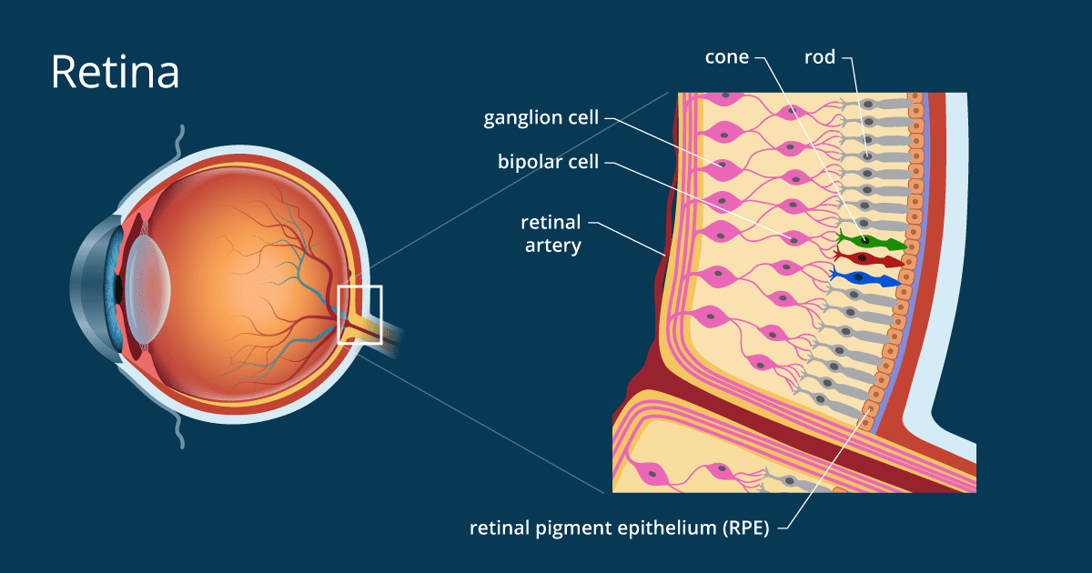
v) Retina:-
The retina is a thin layer of tissue that lines the back of the eye on the inside. It is located near the optic nerve. The purpose of the retina is to receive light that the lens has focused, convert the light into neural signals, and send these signals to the brain for visual recognition.
The retina is the innermost layer of the wall of the eye. It is in immediate contact with the vitreal cavity on one side and with the choroid (of the uveal layer) on the other side. The cellular layers of the retina are as follows:
1) The pigmented epithelium, which is adjacent to the choroid, absorbs light to reduce back reflection of light onto the retina.
2) The photoreceptor layer contains photosensitive outer segments of rods and cones.
3) The outer nuclear layer contains cell bodies of the rods and cones.
4) The outer plexiform layer contains synapses between axons of photoreceptors and dendrites of intermediate neurons.
5) The inner nuclear layer contains cell bodies of intermediate neurons and Muller cells.
6) The inner plexiform layer contains synapses between intermediate neurons and ganglion cells of the optic tract.
7) The ganglion cell layer contains cell bodies of ganglion cells.
8) The optic nerve fiber layer contains axons of ganglion cells. Membrane layers that are not visible in this image separate the photoreceptors from their cell bodies and retina from the vitreal body.
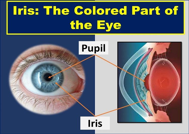
vi) Iris and Pupil:-
Iris:-The colored tissue at the front of the eye that contains the pupil in the center. The iris helps control the size of the pupil to let more or less light into the eye. The iris is the colored part of the eye that controls the amount of light that enters the eye. It is the most visible part of the eye. The iris lies in front of the crystalline lens and separates the anterior chamber train the posterior chamber.
Pupil:-The pupil of the eye is the black circle in the center of the iris. The iris is the colored portion of the eye with a structure and color unique to each person. The pupil of the eye is a portal that admits and regulates the flow of light to the retina. This is part of the process which allows us to perceive images.
The iris and the pupil control how much light to let into the back of the eye. When it is very dark, our pupils are very large, letting in more light. The lens of a camera can focus on objects far away and up close with the help of mirrors and other mechanical devices.
vii) Ciliary body/Ciliary muscle:-
The ciliary muscle is an intrinsic muscle of the eye formed as a ring of smooth muscle in the eye's middle layer (vascular layer). It controls accommodation for viewing objects at varying distances and regulates the flow of aqueous humor into Schlemm's canal.
One function of the ciliary body is to control the lens of the eye. The ciliary body's smooth muscles contract and relax to focus on near or far away objects. Muscle contractions are partly responsible for the round shape of the eye's lenses since fine ligaments directly attach the lens to the ciliary body.
viii) Suspensory ligament:-
Connect the ciliary muscles to the lens and hold the lens in place. (Ciliary muscles, suspensory ligaments lens) Suspensory ligaments pull on the lense; pulls/tighten it. ciliary muscles and Suspensory Ligaments pull on the lens to tighten it. Ciliary Muscles and Suspensory ligaments relax to make the lens go to its natural form.
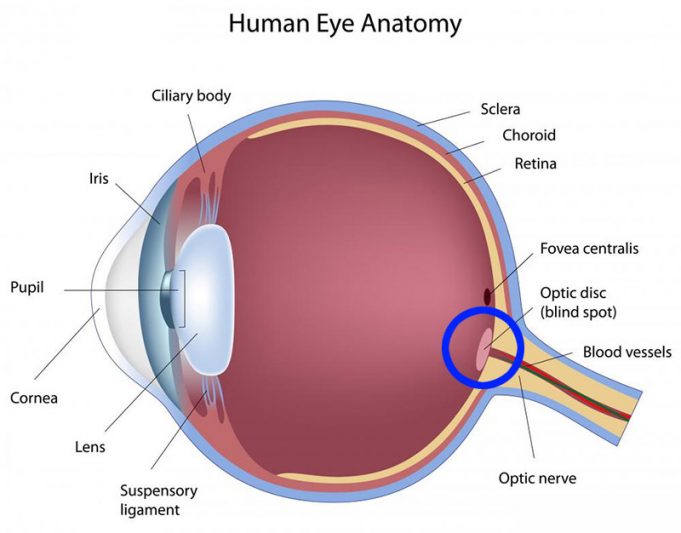
ix) Fovea centralis:-
The fovea centralis, also generally known as the fovea (the term fovea comes from the Latin, meaning pit or pitfall), is a part of the eye, located in the center of the macula region of the retina. The fovea is surrounded by the parafovea belt, and the perifovea outer region. The blind spot (Fovea centralis). This seemingly poor design of the retina, which produces the blind spot in our visual field, is referred to by experts as the inverted eye. The blind spot is located about 15 degrees on the nasal side of the fovea.
x) Optical disc (blindspot):-
Blindspot, a small portion of the visual field of each eye that corresponds to the position of the optic disk (also known as the optic nerve head) within the retina. There are no photoreceptors (i.e., rods or cones) in the optic disk, and, therefore, there is no image detection in this area. When light lands on your retina, it sends electrical bursts through your optic nerve to your brain. Your brain turns the signals into a picture. The spot where your optic nerve connects to your retina has no light-sensitive cells, so you can't see anything there. That's your blind spot.
xi) Blood Vessels and optic nerves:-
Bloodvessels:-Blood supply
There are two circulations of the eye: the retinal (in the retina) and uveal, supplied in humans by posterior ciliary arteries, originating from the ophthalmic artery (a branch of the internal carotid artery).
The optic nerves:- The optic nerve is a bundle of more than 1 million nerve fibers. Also known as the second cranial nerve or cranial nerve II, it is the second of several pairs of cranial nerves. It transmits sensory information for vision in the form of electrical impulses from the eye to the brain.\
This is some information about our eyes.
This is also for the students of class 8/10.
If you want a video in Hindi then see this video.I hope this can give you some knowledge about a body part of our body, that plays an important role in our life.


 The sclera is the white part of your eye. It's a tough, protective covering and the muscles that control eye movement are connected to it.
Comments
Post a Comment
If you have any doubt on any topic please tell me